

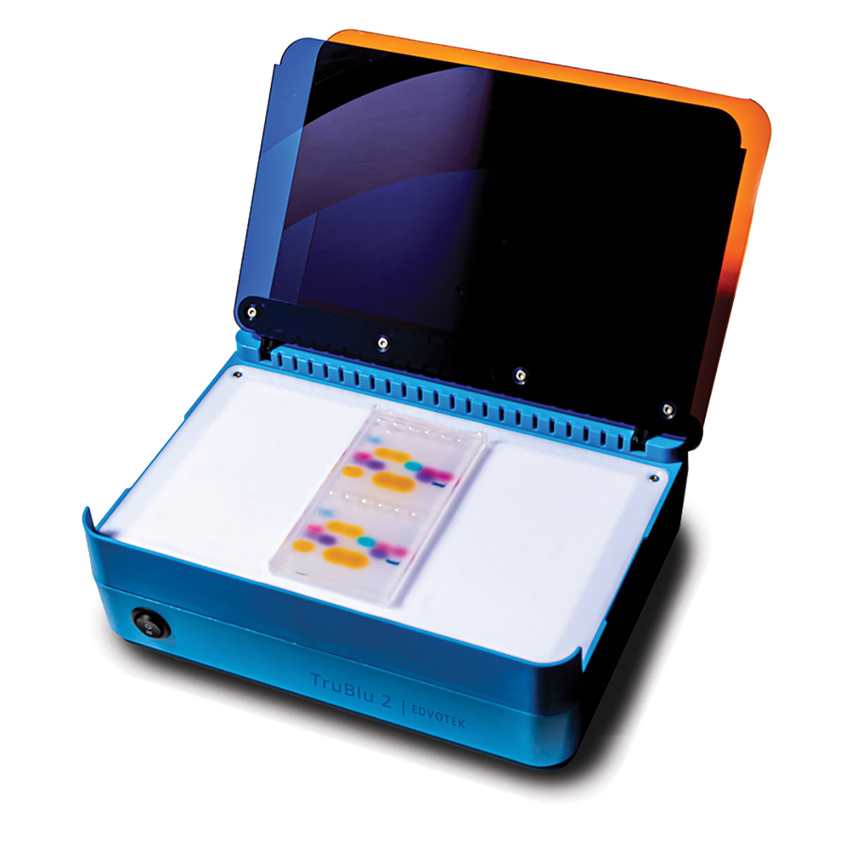
Recently, I had a teacher reach out to me to ask, “What else can I do with the TruBlu2 Transilluminator? It’s a large part of my equipment budget, and I was wondering if it can do more than just looking at gels.” The answer, resoundingly, is YES, the TruBlu2 is incredibly versatile! So, in this blog post, I’m going to cover the many uses of the TruBlu2.
First, we have the TruBlu2’s intended use as a blue light transilluminator for imaging DNA separated using SybrSafe-containing agarose gels. SybrSafe is a classroom-safe fluorescent molecule that specifically binds to and labels double-stranded DNA for visualization. We need to use this stain because DNA is both clear and colorless. Without a DNA stain, we would not be able to visualize the results of our electrophoresis experiment. SybrSafe has been engineered to be less mutagenic and less hazardous than Ethidium Bromide, another common DNA stain. Furthermore, it can be excited to fluoresce with light at 470 nm, eliminating the need for UV transilluminators in the classroom. These innovations help to protect you and your students. (For more about SybrSafe and some FAQ, be sure to check out our previous blog post.)
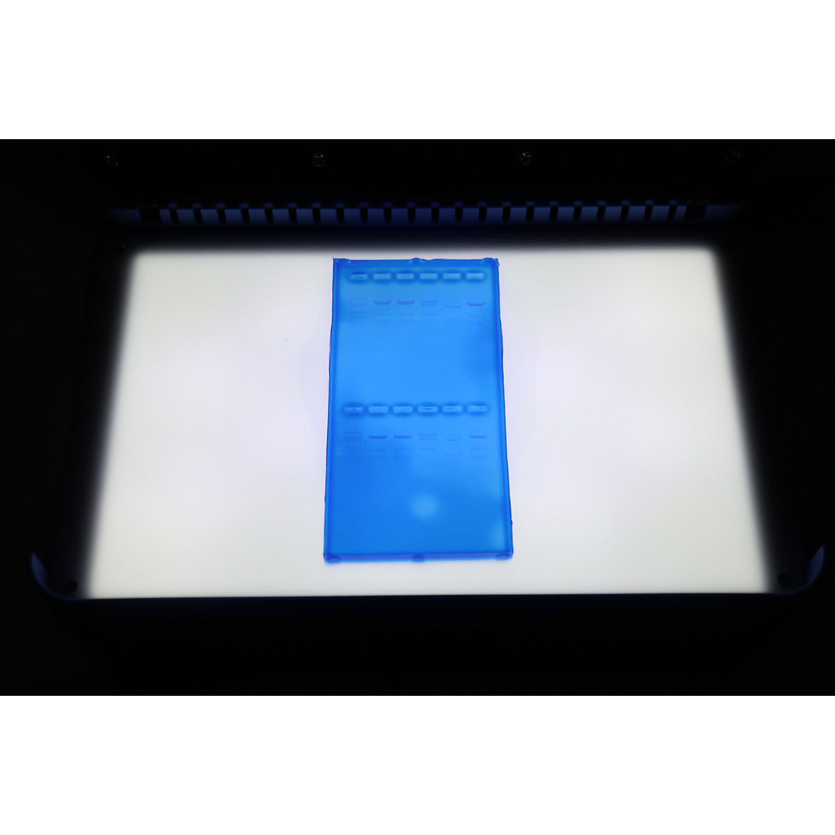
Now, some teachers prefer to use our FlashBlue DNA stain for visualizing the results of their electrophoresis experiments. Other teachers will separate brightly-colored dyes using electrophoresis. The big difference with these experiments is that we do not need to use UV or blue light to visualize the results. Instead, the colors can be seen with the naked eye To enhance the contrast between the bands and the background, we can use the white light feature of the TruBlu2. Simply lift the blue and orange filters and place your gel on the white surface. Using the toggle switch, flip to the white LED setting and view your results! After performing any of these electrophoresis experiments, students can snap a picture with their cell phones to include in their lab reports.

Another use of the TruBlu2 Transilluminator is in visualizing the fluorescence of the Green Fluorescent Protein (or GFP). Like SybrSafe, GFP can be excited by blue light! While we often use UV light to visualize our GFP transformation plates, blue light is just as effective. In fact, in the jellyfish GFP is excited by blue light, which is where the protein was first identified. Therefore, we can visualize the results from our GFP Transformation and our Bioprocessing experiments using the TruBlu2.
So, these applications are taken from experiments that we created for our teachers. But, I was curious… what else could we analyze using the TruBlu2? I spent some time brainstorming, and here are a few things that I discovered could be visualized using a blue light system.
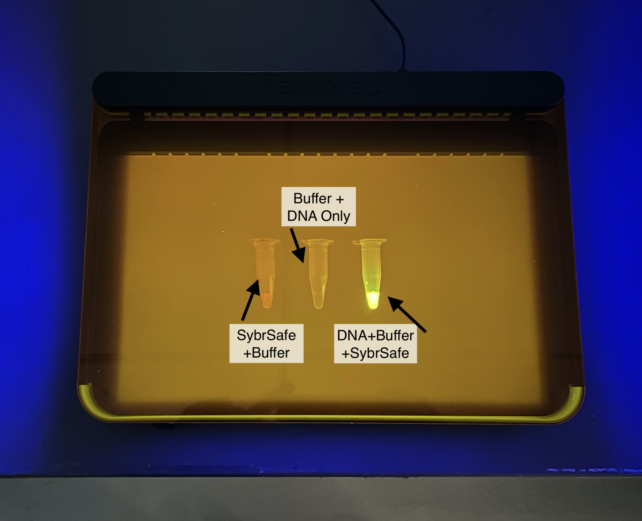
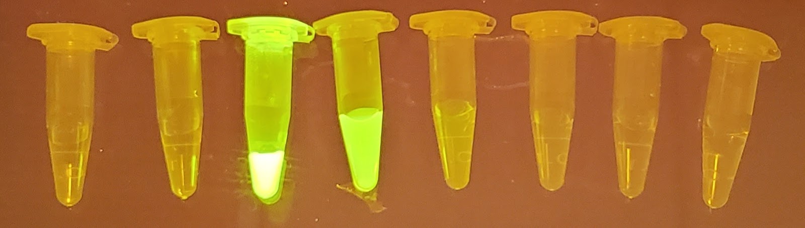
First, could SybrSafe be used to detect the presence of DNA in a buffer sample? From my research, I knew that SybrSafe selectively binds to double stranded DNA. I also knew that its cousin, Sybr Green, is used in quantitative PCR, which simultaneously amplifies and detects targeted DNA allowing scientists to discover the starting amount of a specific DNA sequence in an experimental sample. So, would I be able to use SybrSafe to detect DNA in solution? I set up three tubes: one contained SybrSafe and buffer. The second contained DNA and buffer. The third tube contained SybrSafe, DNA, and buffer. My hypothesis was that I should only observe fluorescence in the third tube. I put the tubes on the TruBlu2 to analyze my results. While a very faint red color could be seen in the SybrSafe + buffer tube, it was obvious which tube contained the DNA, as it glowed with a bright green fluorescence.
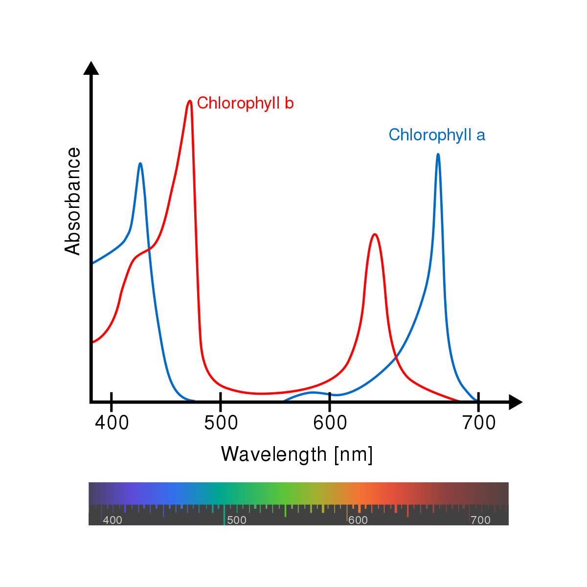
Next, we know that chlorophyll a and b, the plant pigments responsible for absorbing light energy in photosynthesis, absorb light in the blue range. Furthermore, there are other compounds like beta-carotine and xanthophylls which are known to absorb light. So what would happen if we extracted these molecules from plant tissues and visualized them using the TruBlu2? My hypothesis was that we would be able to see the molecules fluorescence.
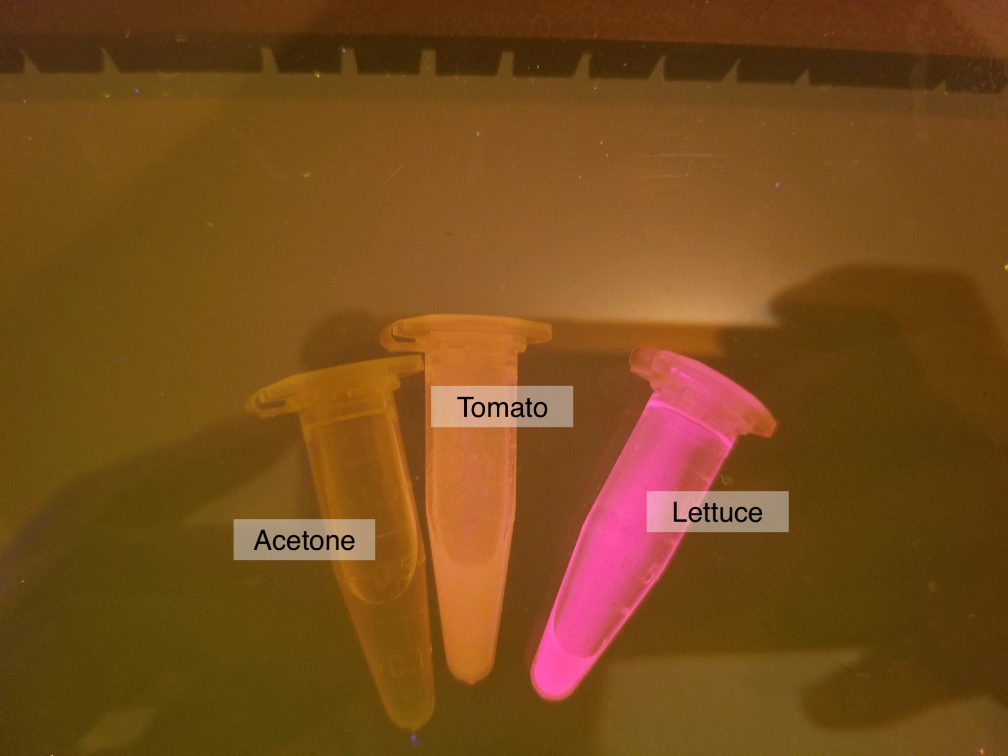
First, I went to the kitchen to grab some vegetables. I had a dark green lettuce, fresh tomatoes, and red cabbage available, so I placed small amounts of each of them in a microcentrifuge tube. As a solvent for my extraction, I added a small amount of acetone (in the form of nail polish remover) to each tube. If you don’t have acetone, rubbing alcohol (i.e. isopropyl alcohol) should also work. I used a chopstick to mash my samples and then transferred the liquid to a second tube, removing it from the mashed veggie debris. I placed the samples on the TruBlu2 and turned it on. To my delight, the tomato and the lettuce fluoresced brightly. The red cabbage had a low reddish fluorescence, which surprised me because the solution was so heavily pigmented I had expected it to be brighter (note — red cabbage not included because my lab partner i.e. my cat knocked the tube off of my desk where it promptly was lost forever, or until I clean under my bookcase). In the future, I’ll save my red cabbage to use as a pH indicator.
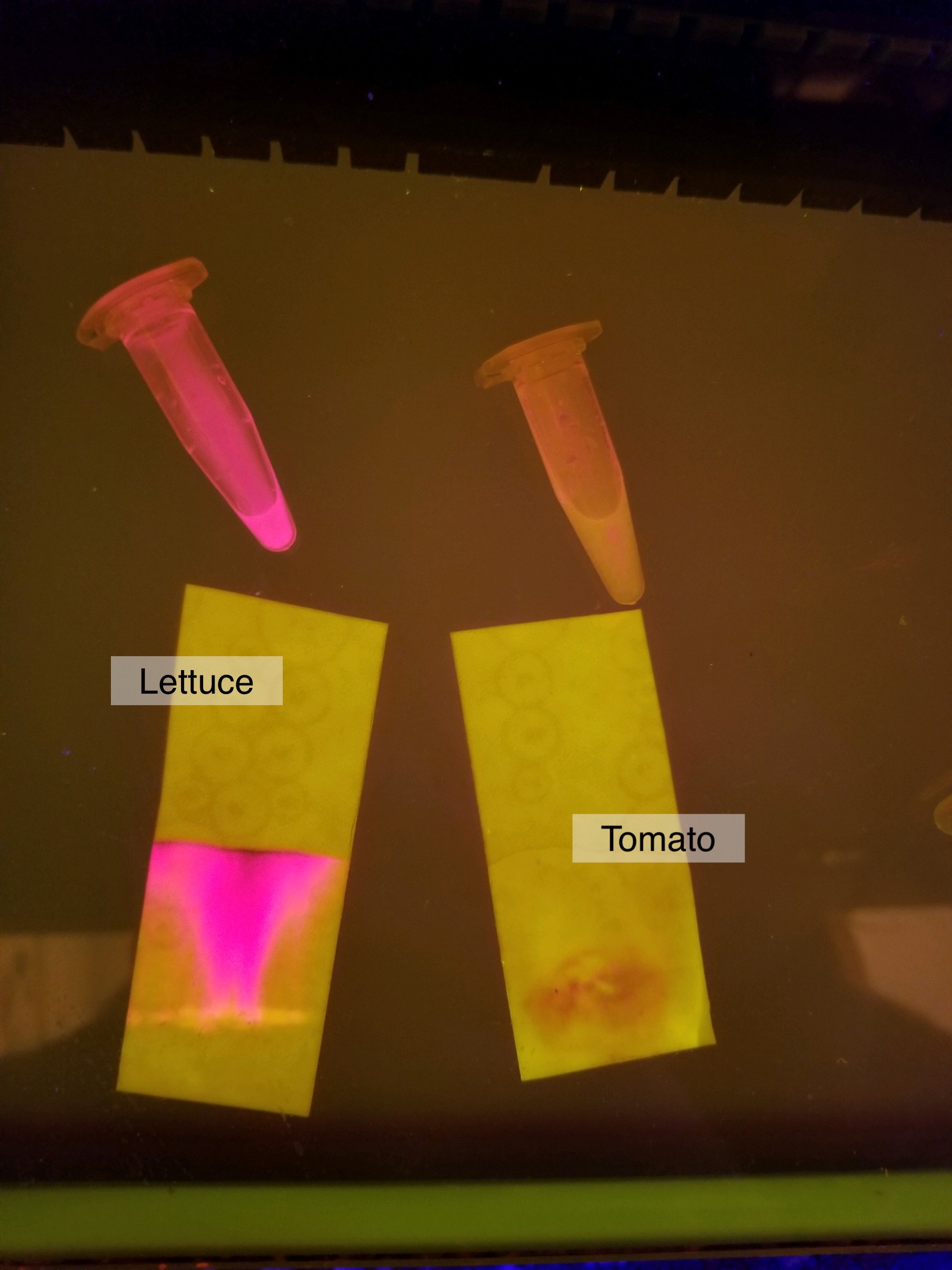
In a previous kitchen chemistry post, we talked about performing chromatography using pigments extracted from plants collected on a nature walk. I wondered whether my pigments, when separated by chromatography, would fluoresce on the TruBlu2. So, I spotted the samples on some filter paper and let them thoroughly dry. Using acetone as my mobile phase, I performed the separation. My hypothesis was that I should be able to see the fluorescent pigments after chromatography, but I was unsure whether the paper would affect this. I was positively gleeful when I saw the pigments fluoresce after the separation, but it’s clear that this technique can use a little research and development to get superior results.
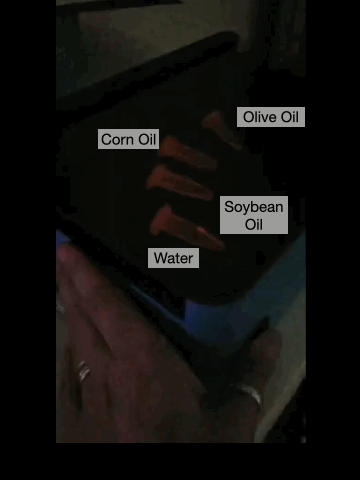
While I was in my kitchen, I realized that I had several plant-based cooking oils: soybean oil, corn oil, and olive oil. I wondered whether any of these oils would fluoresce, which would mean that some chlorophyll or other plant pigment remained in the oil after the plant samples had been macerated and the oils extracted. What I found was that the soybean oil had a very light, yellow glow, while the corn and the olive oil had bright yellow and red fluorescence, respectively. This was very exciting to me in my home-fluorescence adventure!
So, you may ask yourself whether the TruBlu2 a ‘unitasker’? I hope that this blog post showed you that this blue and white light visualization system is a multitasking powerhouse in the biotechnology laboratory! How are you using your TruBlu2 translluminator? Let us know on Twitter, Instagram, or Facebook!

1 comment
Comments are closed.