
Microscopes are amazing pieces of laboratory technology that allow us to visualize objects that are too small to be seen by the naked eye. This includes microscopic creatures like amoeba and bacteria, individual human cells, and even the proteins and organelles within a cell! Microscopes bounce beams of lights or electrons off of the samples. The beams are then amplified using lenses and detected by either the eye (with light) or a detector (with electrons).
Each year, Nikon (a leading microscope company) hosts a science art contest where researchers can send in their favorite microscopic images. The contest, called the Nikon Small World Contest, showcases “the beauty and complexity of life as seen through the light microscope.” Below are our five favorite pictures from the 2021 contest (but be sure to check them all out, they make great teaching resources). Which is your favorite, and why?
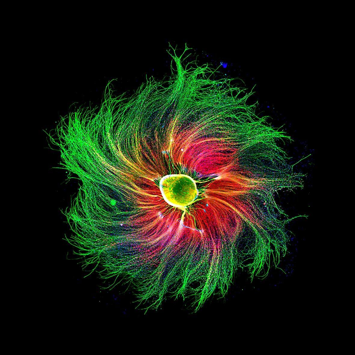
- Sensory neuron from an embryonic rat by Paula Diaz at Pontificia Universidad Católica de Chile
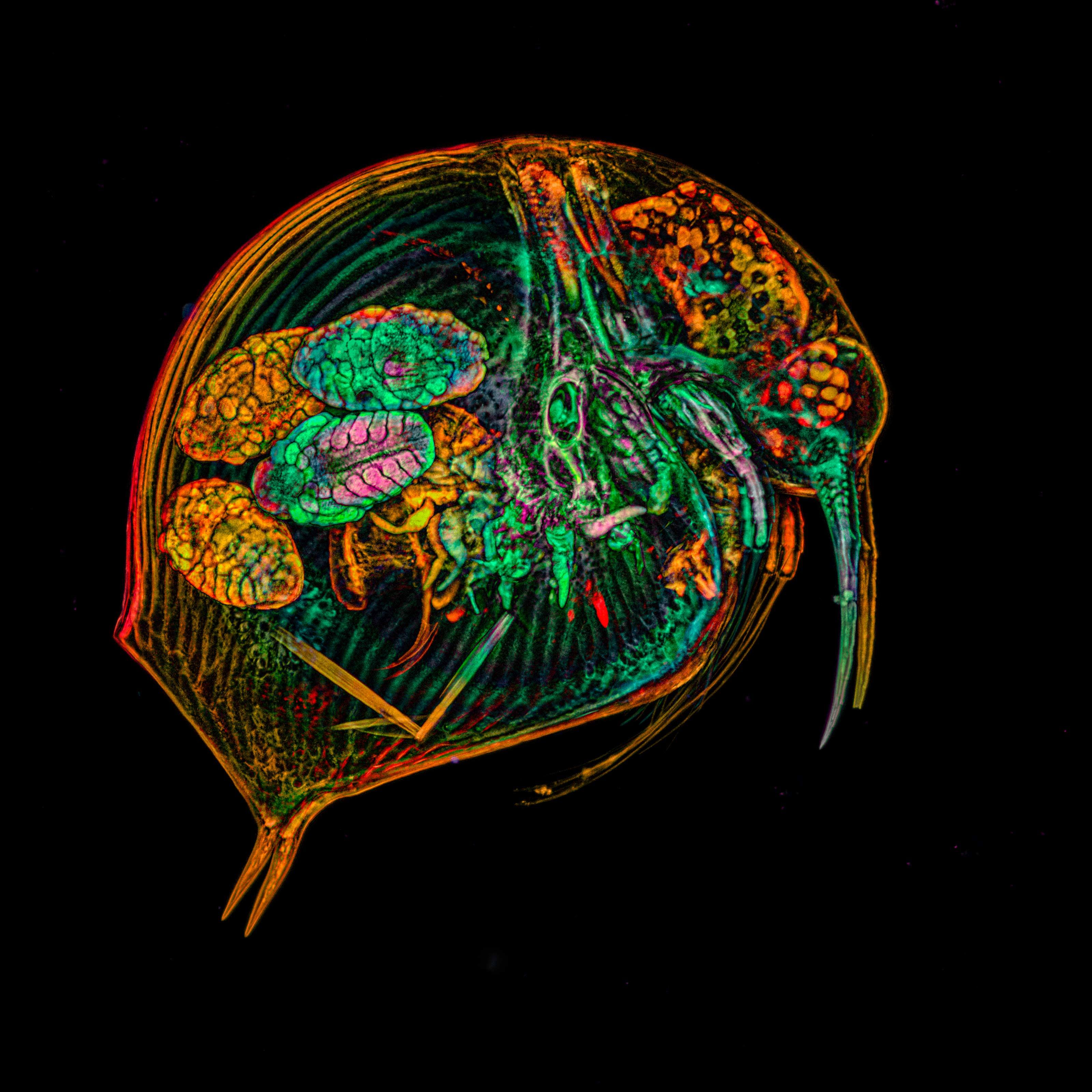
- A freshwater crustacean (Bosmina sp.) by Alexandra Tsitrina at Institute of Developmental Biology Russian Academy of Science
- Oxalate crystals in onion skin by Dr. Robert Markus at University of Nottingham
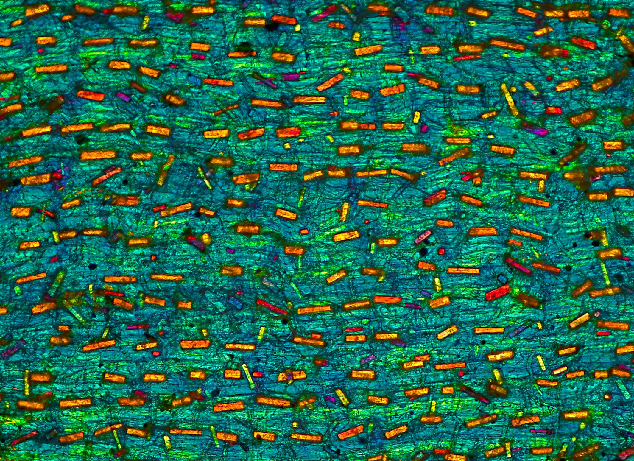
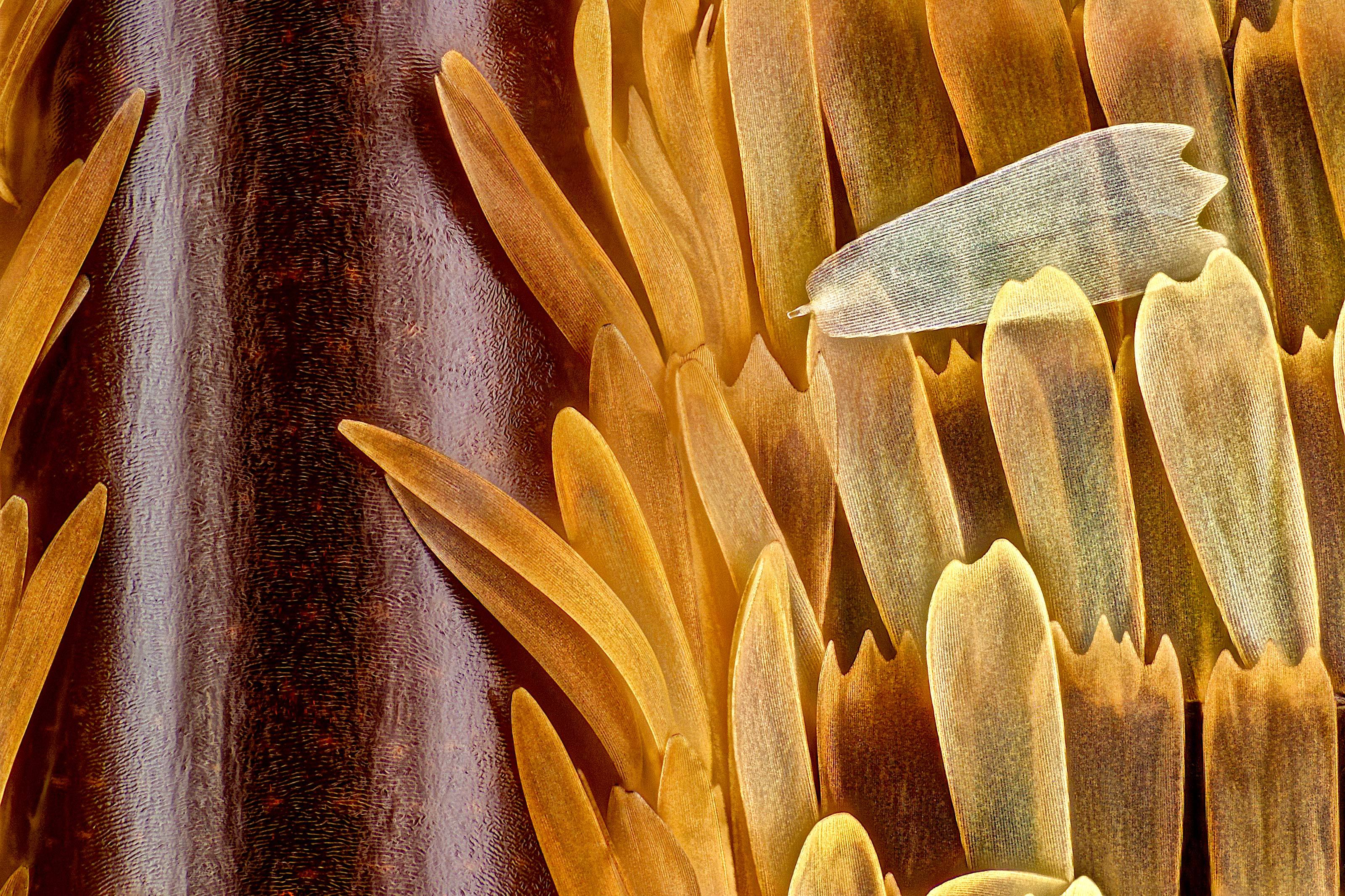
- Vein and scales on a butterfly wing (Morpho didius) by Sébastien Malo at Saint Lys, Haute-Garonne, France
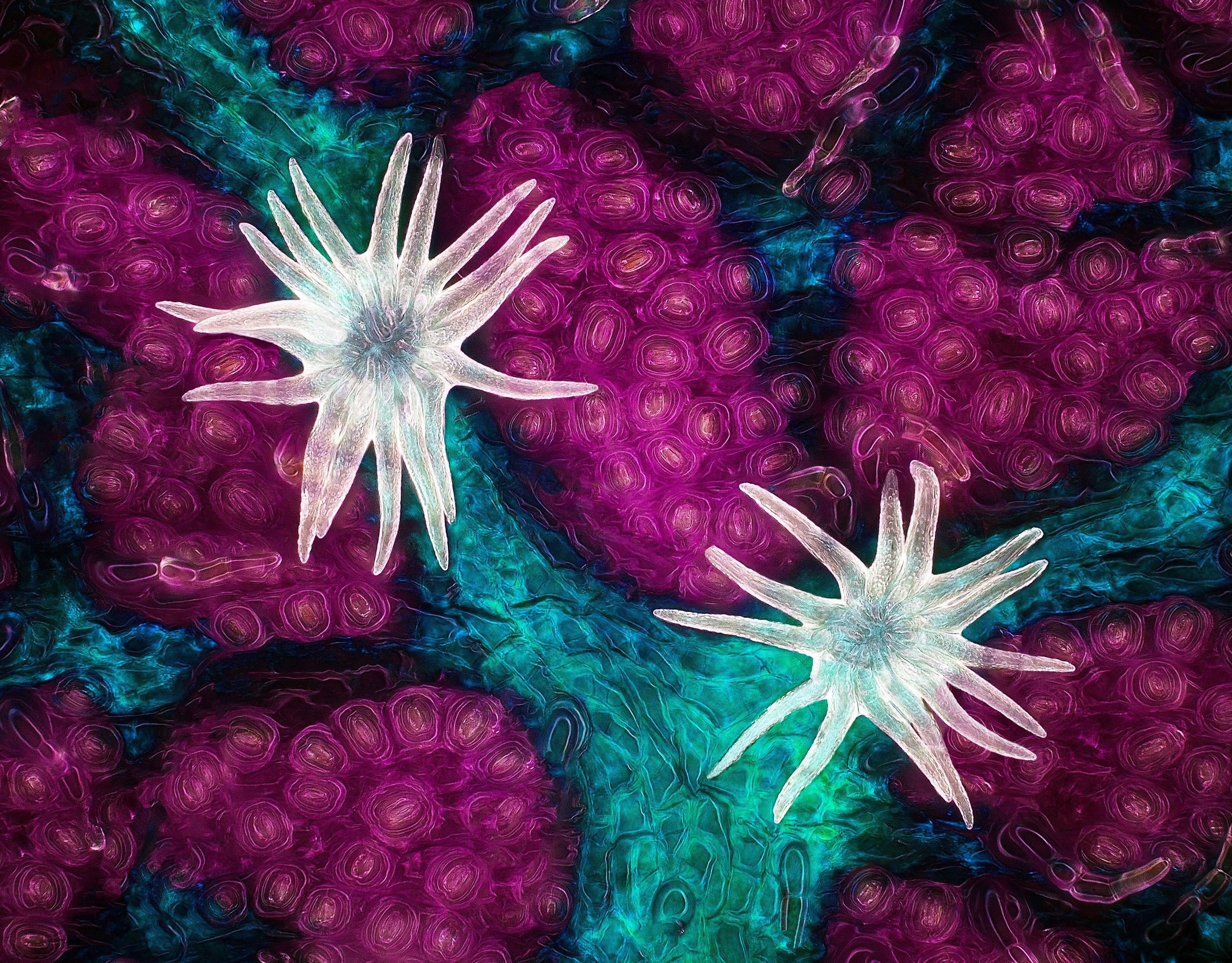
- Trichome (white appendages) and stomata (purple pores) on a southern live oak leaf by Jason Kirk at Baylor College of Medicine.