What is agarose gel electrophoresis?
Electrophoresis is a technique that allows us to separate DNA, RNA or proteins into bands according to their size. For more information about this technique, be sure to check out our previous post describing electrophoresis.
Why do gels need to be stained after electrophoresis?
DNA, RNA, and proteins are colorless in solution. Nucleic acid and protein stains allow us to see the individual bands created by electrophoresis.
What types of DNA stains are used in the laboratory?
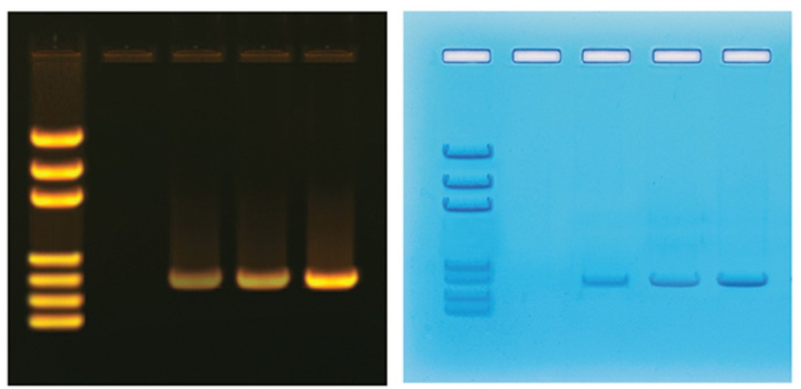
Research laboratories commonly use fluorescent DNA stains because they are extremely sensitive, making it easy to detect small amounts of DNA. However, an ultraviolet (UV) light source must be used to visualize the DNA fragments.
Although visible dye-based DNA stains are less sensitive than fluorescent stains, they are an excellent alternative for the teaching classroom because they are non-toxic. The dye molecules stain the DNA fragments a bright blue color, so special equipment is not required to detect the DNA.
Edvotek ® offers several different methods for visualizing the DNA separated by electrophoresis.
- The easiest and most convenient visible dye-based DNA stain available is InstaStain® Blue. InstaStain® Blue does not require the formulation, storage and disposal of large volumes of liquid stain. Each InstaStain® Blue card contains a small amount of blue DNA stain that is used to visualize the DNA.
- FlashBlue™ is a visible dye-based DNA stain that offers simple and rapid staining of agarose gels. FlashBlue™ is provided as a concentrated liquid stain that, when diluted, can be used for both rapid and overnight staining of DNA fragments.
- The most commonly used fluorescent DNA stain is Ethidium Bromide (EtBr). This molecule binds with the DNA in the gel. When excited with UV light, the EtBr fluoresces and produces a bright orange light. However, because EtBr is a potential mutagen, it must be handled with care. InstaStain® Ethidium Bromide provides the sensitivity of EtBr while minimizing potential contact with hazardous materials by delivering a small amount of stain to the agarose gel via a special paper backing.
- SYBR Safe® is a DNA stain that fluoresces with a bright green color when excited with UV light, similarly to EtBr. Unlike EtBr, SYBR Safe® has been engineered to be less mutagenic, making it much safer to use in the classroom.
How do I use these DNA stains?
Check out our instructional videos at youtube.com/EdvotekInc, or download our Quick Guide to Visualizing DNA for detailed protocols.
What do I do after staining the gels?
 EDVOTEK®’s Midrange UV Transilluminator is designed to visualize DNA stained with either ethidium bromide or SYBR® Safe. The UV filter measures 7 x 14 cm which is optimized for viewing gels cast from EDVOTEK® electrophoresis chambers. Safety features include a UV blocking cover and an automatic power-cut off when the cover is opened.
 The all-new TruBlu™ LED Transilluminator utilizes blue light to view DNA gels stained with SYBR® Safe, thus eliminating the need for UV light or ethidium bromide which can be a hazard in the lab. The spacious viewing area measures 14.5 x 18 cm, which allows you to visualize multiple agarose gels at once. The adjustable control knob allows you to tune the intensity of the light source, ensuring superior visualization results.
 White LED Transilluminator
Our White LED Transilluminator features a spacious 25 x 25 cm viewing area illuminated by long life LEDs and is housed in a slim aluminum body. It’s designed to safely enhance the visualization of DNA stained with FlashBlue™ or InstaStain® Blue.
We hope this helps you get excited about performing electrophoresis in your classroom! Have any other questions about electrophoresis? Please call EDVOTEK’s Technical Support at 1.800.338.6835, available Monday-Friday 8:00am -5pm. We’re happy to help!

1 comment
Comments are closed.